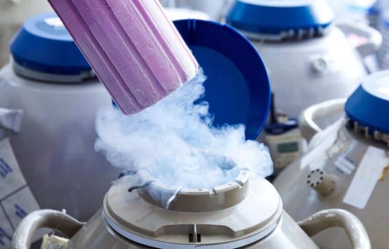
Shiraz Fertility Center Freezing Section
Cryoprese rvation
Embryo Cryopreservation
The first pregnancy resulting from the transfer of a frozen embryo was reported in 1983. In subsequent years, due to advancements in cryobiology, the preservation of embryos in a frozen state has become a fundamental part of modern ART. Embryos can be frozen at any stage, from the zygote to the blastocyst, and these products can maintain their viability for at least several years and perhaps indefinitely.
The cryopreservation process consists of two distinct phases: freezing and thawing. The goal of freezing is to prevent the intracellular water from crystallizing into ice, which would cause cellular damage. The freezing methods vary based on the developmental stage of the embryo, which affects cellular permeability.

There are two primary methods for cryopreserving embryos:
Slow freezing and Vitrification method.
In both methods, cellular water is gradually replaced by cryoprotectants (such as dimethyl sulfoxide and propanediol glycerol) through an osmotic process involving a progressively increasing concentration of the cryoprotectant. In the slow-freezing method, embryos are placed in the freezing medium at room temperature and then transferred to a freezing straw, where they are cooled at a programmed rate of 2°C per minute to -7°C. These samples are then seeded and cooled at a slower rate of 0.3°C per minute to -30°C before final storage in liquid nitrogen (-196°C).
In vitrification, embryos are rapidly frozen by immersion in liquid nitrogen, resulting in a glassy solid state. The process is reversed during thawing. The embryo is gradually passed through decreasing concentrations of cryoprotectant and then incubated in culture medium before transfer. The primary goal of vitrification is to completely eliminate the formation of ice crystals in all solutions containing embryos or oocytes. Most successful vitrification methods rely on the use of very small volumes of sample-containing solutions and direct contact between the embryo or oocyte and liquid nitrogen.

In vitrification, a major concern is the toxic effect of high concentrations of cryoprotectant and the potential risk of cross-contamination from direct exposure to liquid nitrogen. Results of studies have shown that post-thaw survival rates following slow freezing vary between 50 and 90%, and this rate is higher in the zygote stage compared to the cleavage and blastocyst stages. Implantation rates and pregnancy rates after transfer of frozen zygotes, cleavage-stage embryos, and blastocysts using the slow-freezing method have varied in different studies, but this difference has not been significant.
Initial experiences with vitrification have shown that survival rates are consistently high with this method and may result in higher implantation and pregnancy rates. In general, at most centers, the success rate of frozen embryo transfer cycles has been about half to two-thirds of that observed in fresh embryo transfer cycles; one reason suggested is that embryos with the best quality are usually selected for fresh embryo transfer. The advantages of blastocyst freezing over cleavage-stage embryos include better embryo selection during the culture process, which leads to the development of higher-quality embryos compared to frozen cleavage-stage embryos.
One reason for higher pregnancy rates with day 5-6 blastocysts is better synchronization between the endometrial lining of the uterus and the blastocyst. In women with normal ovulatory function, frozen-thawed embryos can be transferred in their natural cycle. Alternatively, embryo transfer can be performed in a stimulated cycle where endometrial development is carefully controlled through the sequential administration of exogenous estrogen and progesterone. Initial down-regulation with GnRH agonists can also be used (as is typically done in oocyte donation recipients). There is no evidence to suggest that one endometrial preparation method is superior to others. In both natural cycles (days after ovulation) and stimulated cycles (days of progesterone treatment), the timing of transfer is coordinated with the developmental stage of the embryo. The advantages of embryo freezing include a reduced risk of multiple pregnancies and reduced frequency of ovarian stimulation.
Oocyte Cryopreservation
Indications for oocyte cryopreservation include women at risk of losing fertility due to the following reasons: malignant diseases requiring pelvic radiotherapy or surgery, ovarian dysfunction (premature ovarian failure, polycystic ovary syndrome, poor ovarian response to stimulation), desire to postpone pregnancy for personal or professional reasons, oocyte donation, legal or ethical concerns regarding embryo freezing, and absence of a male partner’s sperm after successful oocyte retrieval. The reasons for reduced IVF success following fertilization of thawed oocytes include premature release of cortical granules and hardening of the zona pellucida.
The unique nature of the oocyte (large size, spherical shape, singularity, and fragility) is responsible for its low post-thaw survival. In an irregularly shaped cell (such as a fibroblast or lymphocyte), the surface-to-volume ratio is larger, and it reaches osmotic equilibrium more rapidly than an oocyte. Whereas in a large spherical object (like an oocyte), the surface-to-volume ratio is the lowest compared to any other geometric shape. In fact, a smaller surface-to-volume ratio results in a longer duration to reach osmotic equilibrium.
Other complications of freezing include damage to lipid droplets, lipid-rich membranes, and cellular microtubules. Damage to lipids is irreversible and leads to oocyte death. Compared to other species, although the lipid content of human oocytes is relatively lower, damage to the membrane, depolymerization of microtubules, and abnormal chromosome arrangement and aneuploidy may occur following oocyte freezing in humans. During the thawing process and removal of the cryoprotectant, the spherical shape of the oocyte may only allow for limited expansion, and as a result of water influx into the cell, the cell membrane may rupture.
It is best to freeze oocytes between 16 hours after retrieval and immediately after cumulus cell removal. ICSI should be performed approximately 4-2 hours after thawing the oocytes, and maintaining this short culture time is essential to help improve the flexibility of the oocyte membrane (during penetration by the injection pipette during ICSI). The outcome of oocyte freezing depends on the patient’s age, ovarian stimulation regimen, quality of the resulting embryo, timing of embryo transfer, and endometrial preparation method. Ovarian stimulation regimens do not seem to have an effect on the clinical outcomes of frozen oocytes. The main obstacle to the success of oocyte freezing is poor survival, which is due to the size, high water content, and fragile chromosomal arrangement of the oocyte.
During the freezing or thawing process, the formation of intracellular ice damages the meiotic spindle. Another barrier is the hardening of the zona pellucida layer, which hinders natural fertilization. Clinical trial results have shown that oocyte survival after vitrification and warming is 97-90%, fertilization rate is 79-71%, implantation rate is 41-17%, and overall clinical pregnancy rate is 36-61%, with a clinical pregnancy rate per oocyte warmed of 12-4.5%. The survival, fertilization, and pregnancy rates from frozen oocytes are increasing. Although the number of pregnancies and births from frozen oocytes is still low, this number is increasing rapidly. The incidence of chromosomal abnormalities in human embryos derived from frozen oocytes does not differ from those derived from fresh oocytes.
Ovarian Cryopreservation
Recent advancements in cryobiology have brought hope for preserving fertility through the storage and freezing of oocytes and ovarian tissue. Ovarian tissue cryopreservation, at least theoretically, provides a means to freeze primordial follicles for future in vitro maturation. Chemotherapy and radiation therapy in women with cancer jeopardize fertility. In some cases, the ovaries can be moved out of the radiation field. As a method to protect the gonads from the adverse effects of chemotherapy, treatment with GnRH agonists has been proposed, but there is no convincing evidence regarding the efficacy of this treatment.
Currently, autologous ovarian tissue transplantation after cryopreservation seems to be the most practical and effective approach, successfully preserving fertility in women with chemotherapy-induced ovarian failure. In this method, ovarian tissue is surgically removed via laparoscopy or laparotomy and cryopreserved using slow-freezing or vitrification techniques. In the future, the ovarian tissue is thawed and reimplanted into the patient in its original location or nearby (orthotopic transplantation) or in another site such as the forearm or abdominal wall (heterotopic transplantation).
“In orthotopic transplantation, pregnancy may occur spontaneously without the need for assisted reproductive technology, whereas in heterotopic transplantation, in vitro fertilization (IVF) is necessary. Oocytes retrieved from heterotopic grafts in humans have been fertilized in vitro and biochemical pregnancies have been established following embryo transfer. A number of live births have been reported after orthotopic autologous transplantation of frozen ovarian tissue. One of the potential risks of ovarian tissue cryopreservation and autologous transplantation is the reimplantation of tumor cells in women with malignancies. Further research is needed to determine patient selection criteria, tissue collection methods, and cryopreservation techniques.”
